Paramedic
Airway Anatomy
Airway Anatomy
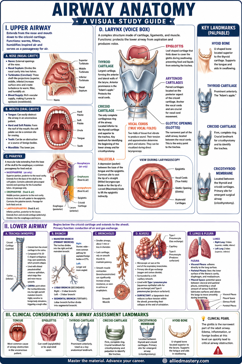
The airway is a critical pathway for the transport of air to and from the lungs. Understanding its anatomy is fundamental for effective airway management.
I. Upper Airway
The upper airway extends from the nose and mouth down to the cricoid cartilage. Its primary functions are to warm, filter, and humidify inspired air, as well as to serve as a passageway for air.
A. Nose (Nasal Cavity)
- Nares: External openings of the nose.
- Nasal Septum: Divides the nasal cavity into two halves.
- Turbinates (Conchae): Three shelf-like projections (superior, middle, inferior) that increase the surface area and create turbulence to warm, filter, and humidify air.
- Blood Supply: Rich vascular supply, making it prone to epistaxis (nosebleeds).
B. Mouth (Oral Cavity)
- Tongue: A large muscular organ that can easily obstruct the airway in an unconscious patient.
- Hard and Soft Palate: Form the roof of the mouth; the soft palate can be a common site for snoring.
- Teeth: Can be an obstruction or a source of foreign bodies.
- Mandible: The lower jaw.
C. Pharynx
- A muscular tube extending from the base of the skull to the esophagus, serving as a common passageway for food and air.
- Nasopharynx:
- Superior portion, posterior to the nasal cavity.
- Extends from the base of the skull to the soft palate.
- Contains the adenoids (pharyngeal tonsils) and openings for the Eustachian tubes.
- Air passage only.
- Oropharynx:
- Middle portion, posterior to the oral cavity.
- Extends from the soft palate to the epiglottis.
- Contains the palatine tonsils.
- Passage for both food and air.
- Laryngopharynx (Hypopharynx):
- Inferior portion, posterior to the larynx.
- Extends from the epiglottis to the esophagus (posteriorly) and cricoid cartilage (anteriorly).
- Divides into the esophagus (posteriorly) and larynx (anteriorly).
- Passage for both food and air.
D. Larynx (Voice Box)
- A complex structure made of cartilage, ligaments, and muscle.
- Function: Protects the lower airway from aspiration and produces voice.
- Key Cartilages:
- Thyroid Cartilage: The largest cartilage, forming the anterior and lateral walls of the larynx. The anterior prominence is the "Adam's apple." It protects the vocal cords.
- Cricoid Cartilage: The only complete cartilaginous ring of the airway. Located inferior to the thyroid cartilage and superior to the trachea. It is a key landmark for identifying the beginning of the lower airway and for cricothyrotomy.
- Epiglottis: A leaf-shaped cartilage located superior to the glottis. It folds down to cover the glottis during swallowing, preventing food and liquids from entering the trachea.
- Arytenoid Cartilages: Paired cartilages located on the posterior aspect of the cricoid cartilage. They anchor the vocal cords and are crucial for vocal cord movement.
- Glottic Opening (Glottis): The narrowest part of the adult airway, located between the vocal cords. This is the entry point to the trachea.
- Vocal Cords: Two folds of tissue that vibrate to produce sound. Their tension and approximation determine pitch and volume. They can be visualized during direct laryngoscopy.
- Vallecula: A depression (pocket) between the base of the tongue and the epiglottis. This is a common place to seat the tip of a straight (Miller) laryngoscope blade or for the tip of a curved (Macintosh) blade to lift the epiglottis indirectly.
II. Lower Airway
The lower airway begins below the cricoid cartilage and extends to the alveoli. Its primary function is the conduction of air and gas exchange.
A. Trachea (Windpipe)
- A cartilaginous tube extending from the cricoid cartilage to the carina.
- Composed of 16-20 C-shaped cartilaginous rings, open posteriorly, which prevent collapse.
- Lined with ciliated pseudostratified columnar epithelium, which helps filter and move mucus upwards.
- Carina: The point at which the trachea bifurcates (divides) into the right and left mainstem bronchi. Highly sensitive to foreign body stimulation, triggering a cough reflex.
B. Bronchi
- Mainstem Bronchi (Primary Bronchi): The trachea divides into the right and left mainstem bronchi.
- Right Mainstem Bronchus: Shorter, wider, and more vertical than the left, making it more common for aspirated foreign bodies or endotracheal tubes to enter the right lung.
- Left Mainstem Bronchus: Longer and more angled.
- Lobar Bronchi (Secondary Bronchi): Each mainstem bronchus divides into lobar bronchi (three on the right, two on the left) to supply the lung lobes.
- Segmental Bronchi (Tertiary Bronchi): Lobar bronchi further divide into segmental bronchi.
C. Bronchioles
- Smaller airways, about 1 mm or less in diameter.
- Lack cartilage, relying on smooth muscle to maintain patency.
- Smooth muscle contraction (bronchoconstriction) can significantly increase airway resistance, as seen in asthma or anaphylaxis.
D. Alveoli
- Microscopic air sacs at the end of the respiratory bronchioles.
- Primary site of gas exchange (oxygen and carbon dioxide).
- Surrounded by a rich capillary network.
- Lined with Type I pneumocytes (squamous epithelial cells for gas exchange) and Type II pneumocytes (produce surfactant).
- Surfactant: A lipoprotein that reduces surface tension within the alveoli, preventing their collapse at the end of exhalation.
E. Lungs and Pleura
- Lungs: Paired organs located in the thoracic cavity.
- Right Lung: Has three lobes (superior, middle, inferior).
- Left Lung: Has two lobes (superior, inferior) to accommodate the heart.
- Pleura: Serous membranes that surround the lungs and line the thoracic cavity.
- Visceral Pleura: Adheres directly to the lung surface.
- Parietal Pleura: Lines the inner surface of the thoracic cavity, diaphragm, and mediastinum.
- Pleural Space: The potential space between the visceral and parietal pleura, containing a small amount of serous fluid that lubricates surfaces and allows the lungs to move smoothly during breathing.
III. Clinical Considerations and Airway Assessment Landmarks
- Tongue: Most common cause of airway obstruction in the unconscious patient.
- Epiglottis: Can swell (epiglottitis) or be aspirated upon.
- Thyroid Cartilage: Prominent anteriorly, used as a landmark.
- Cricoid Cartilage: Firm, complete ring, crucial landmark for cricothyrotomy and palpation to identify the trachea.
- Cricothyroid Membrane: Located between the thyroid and cricoid cartilages; primary site for emergent surgical airway (cricothyrotomy).
- Hyoid Bone: U-shaped bone located superior to the thyroid cartilage, supports the tongue and aids in swallowing. Not part of the larynx itself, but an important superior boundary.
Ready to Go Deeper?
This is just a sample of what Allied Mastery offers. Sign up to unlock our full library, create custom quizzes, track your performance, and more.